
|Articles|May 1, 2008
Oncology NEWS International
- Oncology NEWS International Vol 17 No 5
- Volume 17
- Issue 5
Myoepithelial cells lining milk ducts hold key to spread of DCIS
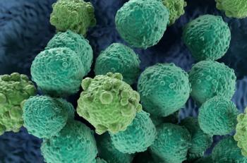
Researchers at Dana-Farber have found that normal myoepithelial cells, which form part of the lining of the milk ducts, suppress fibroblasts that promote tumor growth and invasion, but when certain genes in the myoepithelial layer become under- or overactive, the layer breaks down and disappears, enabling tumor cells to spread.
Advertisement
Researchers at Dana-Farber have found that normal myoepithelial cells, which form part of the lining of the milk ducts, suppress fibroblasts that promote tumor growth and invasion, but when certain genes in the myoepithelial layer become under- or overactive, the layer breaks down and disappears, enabling tumor cells to spread. The abnormal genes include TGF Beta, Hedgehog, and p63. The study was reported in the May 6 issue of Cancer Cell.
Articles in this issue
about 18 years ago
Immunotherapy agent promising in NSCLCabout 18 years ago
NCCN greenlights nilotinib for imatinib-resistant CML patientsabout 18 years ago
No overall survival benefit for dose-intense regimen in SCLCabout 18 years ago
Make a note of new smoking cessation codesabout 18 years ago
Relistor for treating OICabout 18 years ago
Experts argue against need for phase III proton Rx trialsabout 18 years ago
Spotlight on Cancer Centersabout 18 years ago
Novel peptide vaccine promising in myeloid leukemiaabout 18 years ago
Intensive imatinib/chemo ups EFS in pediatric Ph+ ALLAdvertisement
Related Content
Advertisement


Advertisement
Advertisement
Trending on CancerNetwork
1
Investigators Halt Phase 3 Trial of Sacituzumab Govitecan Combo in NSCLC
2
VT-EBV-N Yields Durable Disease-Free Survival in Pretreated ENKTL
3
Melanoma Trials You May Have Missed at ASCO 2026
4
ASCO 2026: Translating the Top Prostate Cancer Data Into Clinical Action
5