|Articles|February 2, 2023
- ONCOLOGY® Companion, Volume 37, Supplement 1
- Volume 37
- Issue 1
- Pages: 6-9
The Genetics of Multiple Myeloma: Expert Perspectives
Author(s)ONCOLOGY Staff
Experts discuss the genetics of multiple myeloma, the prevalent testing paradigms in this space, and possibilities for the future.
Advertisement
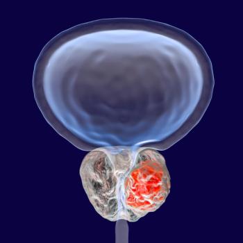
At an Around the Practice program hosted by CancerNetwork®, a panel of experts discussed the pathogenetic and cytogenetic characteristics of multiple myeloma, as well as the prevalent testing paradigms in this space and possibilities for the future. The discussion was led by Joshua Richter, MD, an associate professor of medicine at the Tisch Cancer Institute and director of multiple myeloma at the Blavatnik Family Chelsea Medical Center at Mount Sinai in New York, New York.
Panelists included Karthik Ganapathi, MD, PhD, an associate professor of pathology in the Department of Laboratory Medicine and attending hematopathologist at the University of California, San Francisco; and Adrian M. Dubuc, PhD, an assistant professor of pathology and assistant director of the cytogenetics lab at Brigham & Women’s Hospital Center for Advanced Molecular Diagnostics at Harvard Medical School in Boston, Massachusetts.
Cytogenetic Testing
RICHTER: What test do you conduct in patients with a suspected diagnosis of multiple myeloma?
DUBUC: Cytogenetics remains crucial. It [provides] so much diagnostic and prognostic information in many hematologic malignancies, including myeloma.
There’s been a long history of using karyotype analysis: an unbiased genome-wide assessment of the patient’s karyotype [to determine] several structural variations that could occur across the genome. Traditional karyotypic analysis remains a poor way to detect abnormalities in myeloma. In many studies, fewer than 30% of patients have an abnormal karyotype.
To enhance our ability to detect abnormalities, we try to stimulate myeloma cells with mitogens like IL-4. Typically, [these cells] sit in culture for 4 to 5 days, 5 days being the standard. But even with those best practices developed and espoused by the American College of Medical Genetics and Genomics, the detection rate remains poor.
Therefore, we rely primarily on fluorescence in situ hybridization [FISH] to confirm or refute the presence of crucial pathognomonic aberrations. FISH is often coupled with plasma cell enrichment. It’s currently important to try to enrich the nuclei we score from myeloma cells, or from plasma cells in general. This increases the chance of detection.
FISH and karyotyping—FISH in a plasma cell–enriched capacity—remain the primary modalities of cytogenetic study. There are emerging next-generation sequencing technologies, but they’re not presently used in the vast majority of clinical analyses we perform.
RICHTER: What are some of the typical cytogenetic alterations seen in patients with myeloma, and how do they contribute to pathogenesis? Which are considered primary events, and which are considered secondary?
GANAPATHI: Essentially all patients with multiple myeloma have precursor lesions, often referred to as monoclonal gammopathy of undetermined significance [MGUS], and a smoldering disease process before they become overtly symptomatic. Just like [with] other hematologic malignancies, we now know that multiple myeloma and other plasma cell neoplasms are associated with certain recurrent genetic abnormalities that drive the pathogenesis and biology of this disease.
We can divide cytogenetic abnormalities in plasma cell neoplasms into 2 categories: the early lesions—those genetic abnormalities which occur early in the MGUS or smoldering myeloma stage—and the secondary abnormalities that usually occur [later].
The early abnormalities in plasma cell myelomas can themselves be divided into 2 subcategories. One is copy abnormalities, usually called hyperdiploidy, which are increases in the odd-numbered chromosomes in the myeloma clone, except chromosomes 13 and 17. These occur in about 40% of all patients [with myeloma]. The other subcategory, containing about 55% to 60% of patients, comprises abnormalities involving rearrangements of the immunoglobulin heavy chain [IgH], which affect chromosome 14q32. There are 5 genes that are typically rearranged within the IgH: FGFR3 or MMSET, now referred to as NSD2, on chromosome 4b of the gene cyclin B3; CC and B3 on chromosome 6b of the gene cyclin B1 on chromosome 11q; the gene map on chromosome 16q; and the gene map B on chromosome 20q. These 5 are the classic genes that undergo rearrangements.
Moving on, the secondary abnormalities are often overlaid on a background of these primary abnormalities, and the more common include abnormalities of chromosome 1—specifically, a loss of the short arm of [chromosome] 1p and a gain of the long arm of chromosome 1q. That’s a common secondary abnormality. The 2 other common [secondary] abnormalities are deletion of the long arm of chromosome 13—[a condition called] monosomy 13q—and 17p deletion. These secondary abnormalities often occur in the setting of primary abnormalities.
RICHTER: How do you approach conventional karyotyping in FISH?
GANAPATHI: Interestingly, plasma cells are quite fragile and don’t survive well outside the body, unlike, say, leukemic blasts. We’re [therefore] at a disadvantage when trying to grow these cells [in cultures]. As Dr Dubuc said, they need stimulants, and usually a few days to grow and divide, to reveal these cytogenic abnormalities. From a patient care point of view, the long wait time is not ideal.
We often use FISH on nondividing cells to get a genetic snapshot much faster and thereby obtain a complete diagnostic report. [As] plasma cells do not survive well outside the body, examining a nonselected pool of bone marrow cells to perform FISH is not ideal. The [diagnostic] yield of plasma cells is often much lower than it would be in a bone marrow biopsy. The sensitivity of nonselected FISH testing is quite low in plasma cell myeloma, so to circumvent the low yield of plasma cells, we often use a technique where we enrich a few plasma cells from the bone marrow aspirate sample and then perform a FISH test. This enrichment significantly improves diagnostic yield.
RICHTER: Do physicians routinely order cytogenetic testing for these patients in practice?
DUBUC: I use the term “cytogenetics” to mean any test that detects either numerical or structural aberrations of the genome. Cytogenetics is often associated with karyotyping, the predominant testing modality. At our practice here in Boston, almost all patients with a presumed or confirmed myeloma diagnosis undergo FISH-based testing. We perform a karyotype in very limited instances because it provides such a low yield of accurate and reliable abnormality detection.
It’s important to note that a lack of detected abnormalities in karyotype-based analyses doesn’t mean there won’t be a result issued [to the patient]. It’s very common for patients with myeloma to return a normal result, which could very likely represent a false negative. That normal karyotype [could simply be] reflecting the non-neoplastic component of the specimen, and [therefore] isn’t an accurate depiction of the underlying cytogenetic abnormalities present in the specimen. Largely because of this trend, we rely on FISH-based studies to evaluate the totality of the aberrations we can detect. The challenge here is that we often use a limited number of plasma cells [that] we’re able to isolate through a plasma cell–enrichment process. We often use a tier-based methodology, in which we start [by examining] the high-risk [epigenetic] markers, [specifically] chromosome 17p alterations affecting 1p or 1q—the presence or absence of an IgH tier arrangement. Using those 3 tests as a tier-1 test, we can then gauge the need for additional operations. If there is evidence of an IgH tier arrangement, we can reflex more broadly to a panel of FISH-based tests to identify the specific IgH partner. In the absence of an IgH tier arrangement, that’s not necessary. We try to complete as comprehensive a FISH panel as possible, particularly for newly diagnosed patients because new diagnoses include all the high-risk markers as well as the presence or absence of all the identifiable IgH pre-arrangement partners.
GANAPATHI: Here at the UCSF, we perform conventional karyotyping in addition to FISH at diagnosis for myeloma-specific abnormalities. One benefit of conventional karyotyping is that, in a small subset of patients with any class of neoplasms, they can have other [additional] myeloid neoplasms, such as Nijmegen breakage syndrome, which can have some regular cytogenetic abnormalities that are picked up by conventional karyotyping. [Conventional karyotyping] is certainly important in those patients. For a few, it may alter their subsequent management and therapy options.
RICHTER: When should clinicians repeat these tests, if ever?
GANAPATHI: Clinicians like to have the complete picture at diagnosis, [before] mapping out potential treatment plans for these patients. We certainly would like to get a FISH [score] upon a diagnosis of myeloma, and [name] the disease [as such] so the labs will know what [they’re working with]. That’s our normal protocol. The way in which the diagnosis and disease [develop] will [determine whether we repeat tests].
RICHTER: What are the enrichment processes at your labs? How long does it typically take?
DUBUC: We have largely moved away from curative analysis. The yield for apparitions associated with plasma cells is low. It can be critical to identify secondary apparitions. Often, in unstimulated curative [environments], we set up multiple cultures with different mitogens to identify abnormalities in cell images, with the goal of protecting any apparitions present within the myeloma cells.
When a bone marrow sample arrives at our lab, a technologist will take the specimen through a magnet feed–based selection process, ideally within 24 to 48 hours. This is designed to identify or enrich cells that have CD138 expression. The timeframe is incredibly critical; it’s well established that plasma cells can undergo CD138 shedding. A sample that sits over a weekend—or any length of time beyond 72 hours—will potentially have a decreased enrichment capacity. [Thus], a sample will ideally move through the lab as quickly as possible.
We select positively for those cells that are CD138-enriched. At that point, we drop them in as many wells as we can, inside which we can quickly profile them with FISH probes and look for those crucial abnormalities. The whole process might only take a few hours from the time of arrival to the time we drop cells and begin applying a FISH-based probe. The time frame depends partly on the specifics of the required incubation and on the various cycles of washing that occur. It’s a challenge to maximize both speed and efficacy [given] how many samples [tend to] arrive at the same time. [However], we’re very fortunate to have a large myeloma clinic here, [which allows] us to process in parallel. We have a single, dedicated group of technologists who undertake this process again and again to ensure efficient enrichments.
Past and Present Developments
RICHTER: What are some of the data supporting the use of plasma cell enrichment to detect these genetic abnormalities in myeloma?
GANAPATHI: The fluorescence-activated and magnetic-activated cell sorting techniques exist to purify plasma cells. These have been studied for at least the last 13 years, and [data from] initial studies showed a significant improvement in abnormality detection in plasma cell myeloma [using these methods]. Most studies examined the percentage of plasma cells obtained pre-enrichment, or unenriched, vs post enrichment, and the data are staggering. You can have a 3- to 74-fold increase in plasma cell yield in enriched vs unenriched samples.1 [Although] that number seems hardly believable, [the data are] fascinating. This is an orders of magnitude increase in yield using magnetic or flow sorting. The sensitivity of FISH testing is approximately 8% in unenriched plasma cell myeloma samples vs 72.5% in samples enriched for plasma cells. The detection rate of abnormal cytogenetics at diagnosis, which is such a key factor when issuing diagnoses, is many orders of magnitude better in the enriched sample.
The evidence is stark: Plasma cell enrichment is the preferred way to detect abnormalities in myeloma.
When testing follow-up samples, the plasma cell burden is often not very high. At diagnosis, a sample from a patient can comprise 60% to 70% plasma cells, which makes both the enrichment and the acquisition of positive results straightforward. Follow-up samples meanwhile might comprise less than 5% plasma cells. Even on the diagnostic bone marrow specimens I examine as a hemopathologist, my limited detection methods may not be enough to find the residual disease.
On the other hand, if you perform magnetic sorting or flow cytometry–based sorting and FISH on these specimens, which morphologically appear to be negative marrows, you can successfully pick out clones, whether they be existing clones or new abnormalities. So these tests can work in both settings, both at diagnosis and in follow-up specimens.
DUBUC: It’s also important to note 2 other variables [that affect] the sensitivity of FISH-based testing. Firstly, individual cytogenetic and diagnostic laboratories will establish their own cutoffs for positivity vs negativity for each of the probes used across every disease, including myeloma. FISH can often detect abnormalities only at the level of 1% or greater. The capacity of FISH to detect those abnormalities in the absence of enrichment is incredibly poor. So again, enriching the population [helps ensure] the vast majority of the nuclei we evaluate are representative of the specific cell type that gives rise to this disease. Enrichment helps us more accurately detect the presence or absence of abnormalities.
RICHTER: What are some of the key barriers to the widespread adoption of these practices?
DUBUC: Historically, [many facilities have faced persistent] challenges trying to increase clinical nuance by offering the totality of the tests they feel are important. Currently, from a community perspective, it’s critical for us to refuse to accept substandard results. We have to push ourselves as diagnosticians to ensure we’re offering the highest-quality results. Part of this requires accurate verifications of the types of atlases we perform.
Another barrier to some groups, including our own, has been [overcome by] working alongside our colleagues in pathology and oncology to ensure we receive [timely] specimens to perform accurate validation studies. This way, we can offer standard-of-care testing and also continually improve [our tools and methods]. For example, if we can’t perform enrichment, what are the steps to [circumvent] that problem?
Ideally, we can use samples to validate the effectiveness not only of the enrichment itself [but of our ability to] consistently achieve enrichment, and [at the same time] maintain the capacity to return FISH results which are, in some ways, in line with parallel studies. This can be challenging for some based on the volume of myeloma samples they receive. Fortunately, among the cytogenetics community, there’s a very collegial attitude toward sharing best practices. [We try to] learn from the success and failures of others and use them to design the most efficient and effective validation studies possible. [We do all of this] to ensure we can issue timely, accurate results to our clinical colleagues.
GANAPATHI: Additionally, it’s critical for smaller hospitals, which might not have the bandwidth or patient volume to sustain a fresh myeloma [specimen], to send patient samples to reference labs to get testing done with haste. Unlike in lymphoma, in which genetic tests can be performed retrospectively on older samples, testing must be performed on fresh samples in myeloma, and bone marrow specimens are the freshest. Clinicians must do this when the [normal] procedures are finished. You’ll miss that [small] window of opportunity [otherwise].
Future Directions
RICHTER: How might we use cytogenetics to further classify myeloma into more than just a single disease?
GANAPATHI: The new International Consensus Classification follows up on the old World Health Organization classification, which divided multiple myeloma into distinct genetic subcategories [based on the] common cytogenetic abnormalities [such as]: multiple myeloma with hyperdiploidy; multiple myeloma with cyclin D1 rearrangement, including of cyclin D1/11q and cyclin D1/11p; multiple myeloma with NSD2 translocation, which is the multiple myeloma SET domain [MMSET] FGFR3 aberration; and multiple myeloma with MAF gene rearrangements.2 These subcategories are baked into the new myeloma classification.
From a genetic standpoint, myeloma classification is moving to where AML classification was 15 or 20 years ago when genetic rearrangements became independent subcategories. This is going to drive reclassification of this disease and allow more personalized therapy. These [subcategories] will also open patients to potential trials of the new therapeutics we can offer.
DUBUC: As a cytogeneticist, hearing that so many things in this disease are increasingly reliant on our ability [to determine the] cytogenetics puts a smile on my face. We’ve seen this evolution manifest in other disease types, and it’s nice to see it in myeloma.
RICHTER: Are there any other developments on the horizon?
DUBUC: I can see 2 immediate paths [for improvement] moving forward. One is to ensure we consistently achieve the appropriate diagnosis using a combination of [plasma cell] enrichment and accurate assessment of the critical [cytogenetic] aberrations. [The other is that] we know there are certain types of genetic alterations that can result in false-negative FISH results, so we must continue to push forward, using molecular tools and novel algorithms, [for example], to further dissect these diseases and better understand their clinical heterogeneity.
There’s still so much more to learn, and [although] there’s an incredible benefit to rapid and targeted testing with FISH, we lose some capacity to appreciate the genome-wide structure and nature that can occur [in the process]. There’s still considerable work to be done in understanding the additional genomic alterations that occur.
Final Thoughts
GANAPATHI: We’ve seen incredible advancements in myeloma diagnostics. Official classification of myeloma based on lycorine cytogenetic abnormalities will be the platform upon which all prospective trials will be designed, which will be the springboard to examining future molecular aberrations in GS-based mutation analysis. This is where the AML and the lymphoma worlds have led us. Myeloma is well on the way, from a pathological and diagnostic standpoint, toward more accurate subclassification, [allowing] tailored therapeutics and prognostics.
DUBUC: There’s so much value in these kinds of conversations. Bringing together the worlds of oncology, pathology, and genomics [is always beneficial]. In having these conversations, we can accurately assess where we are and where we need to go.
RICHTER: Here are my 2 cents. It has been demonstrated from a clinical standpoint that patients who are evaluated at an academic myeloma center like any of ours at least once will experience better outcomes than those whose care is carried out solely within a community setting. Your bone marrow is important [enough] to demand those second opinions and [improved] care. Of course, the day-to-day chemotherapy can be administered in a community setting with perfect expertise, but it’s clear that bone marrow [samples] should be evaluated in high-level labs [for maximal efficacy], at least for now.
It’s necessary for all of us to collaborate when plotting the optimal course of treatment for patients.
References
- Lu G, Muddasani R, Orlowski RZ, et al. Plasma cell enrichment enhances detection of high-risk cytogenomic abnormalities by fluorescence in situ hybridization and improves risk stratification of patients with plasma cell neoplasms. Arch Pathol Lab Med. 2013;137(5):625-631. doi:10.5858/arpa.2012-0209-OA
- Campo E, Jaffe ES, Cook JR, et al. The International Consensus Classification of mature lymphoid neoplasms: a report from the Clinical Advisory Committee. Blood. 2022;140(11):1229-1253. doi:10.1182/blood.2022015851
Articles in this issue
Advertisement
Related Content
Advertisement


Advertisement
Advertisement
Trending on CancerNetwork
1
Study Redefines Functional High-Risk Multiple Myeloma in Modern-Therapy Era
2
Half a Year Into the Breast Biopsy Needle Shortage, Clinicians Lean on Networks, Not Workarounds
3
Acupuncture Combo Improves Quality of Life in Head and Neck Cancer
4
Resistance Biology: MARIPOSA vs. FLAURA2 Reshaping Debate
5