Ayal Aizer, MD, MHS, discussed Alliance A071801 trial data favoring fractionated SRS over single-fraction SRS after brain metastasis resection.
Josef Vymazal, MD, DSc, discusses a prospective study comparing delayed single- and double-dose gadopiclenol MRI for detecting small brain metastases.
At the 2026 SNO ASCO CNS Metastases Conference, Josef Vymazal, MD, DSc, presented a prospective, single-center study that compared delayed single-dose and delayed double-dose gadopiclenol (Elucirem) MRI for detecting brain metastases prior to stereotactic radiosurgery. In the study, 156 patients underwent standard postcontrast imaging with the recommended dose of gadopiclenol at 0.05 mmol/kg, followed by delayed imaging roughly 13 minutes later. Half of the patients received an additional dose of gadopiclenol before delayed imaging (delayed double dose), while the rest did not (delayed single dose).
The delayed double-dose imaging detected significantly more consensus lesions than standard non-delayed imaging, while delayed single-dose imaging did not. The detection advantage was driven predominantly by small lesions, with 79% of additional lesions detected on delayed double-dose scans measuring 5 mm or smaller.
Vymazal discussed the clinical rationale for the study, including why not all MRI contrast agents perform equally for this purpose, where the 13-minute delay in the protocol originated, and what the differential detection advantage of the double-dose approach suggests about the underlying pharmacokinetics of gadopiclenol. He also addressed the logistical and financial implications of adopting this protocol.
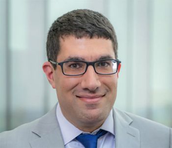
Vymazal is the head of the Department of Radiology and deputy director for Science and Research at Faculty Hospital Motol and Homolka, and a full professor at Charles University in Prague, Czech Republic.
Vymazal: Currently, with Gamma Knife or CyberKnife radiosurgery, we can treat 10 or even more metastatic lesions. In the past, we treated fewer lesions, and we missed some lesions. Right now, we want to see as many lesions as possible because we can treat 10 to 12 metastatic lesions of a really very small diameter. This is the reason that we try to image: to visualize as many metastatic lesions as possible, which does not always happen in routine clinical practice.
It is important to bear in mind that MRI contrast agents are not equal. There is even more difference than [there is] in CT, and this is very important not only for radiologists but also for neuro-oncologists and neurosurgeons. There is a huge difference between individual contrast agents. In layman’s language, some of these contrast agents are very weak, and some of them are very strong. If the contrast agent is weak, it does not mean that it’s a bad contrast agent. It can be cheap, it can be very safe, and for detecting benign brain tumors like meningiomas or schwannomas, I really don’t care if the final image is a little bit grayer or a little bit brighter. For detecting small enhancing lesions, which [can come from non-breast metastases sources]…it is very important to use contrast agents that are strong. They have high relaxivity. We have been working with a contrast agent that has the highest relaxivity on the market, so we assume that we will be able to detect the smallest metastatic regions with this contrast agent. This contrast agent is so strong, so powerful, that it is approved at half dose compared to other macrocyclic or other contrast agents that are used in clinical practice.
We designed our study such that we [randomly assigned patients to receive] a single dose, which is the recommended half dose…or the double dose—which is, in fact, not a double dose, but a standard dose for other contrast agents. The question was: did we detect more metastatic lesions with the double dose, in comparison with the half dose of this contrast agent? The result was statistically significant. We scanned 159 patients, divided randomly into 2 groups, and there was a statistical increase in detecting small metastatic lesions with the double dose…. The increase is quite significant; in every second patient, statistically speaking, [there was] one more lesion.
We also used other sequences—we presented only some of the imaging sequences, we also used the whole imaging protocol. There were earlier studies with a delayed single dose, where they applied the contrast agent and then waited 13 minutes—between 10 and 15 minutes—and the diagnostic yield was better. We wanted to also use this schedule. That’s why there was this delay.
That the pharmacokinetics of this contrast agent are probably a little bit different from other contrast agents. The increase in contrast enhancement after the application of the contrast agent is probably faster in comparison with other contrast agents. Practically, it tells us that if the patient signs a special informed consent form for off-label use of a double dose of the contrast agent, the patient may benefit from the exam because we are able to detect more lesions. The benefit for the patient in this case is more important than the limited, higher burden of gadolinium for the patient who really needs to have all brain diseases treated.
This is exactly the point, and it is a very important point. If the lesion is bigger, you see it even with the weak contrast agent, or with a single dose—maybe it is not so bright—but with small lesions, about 2 to 3 mm in diameter, you can completely miss it with the lower dose of the contrast agent. This is because of the size of the lesion.
It would be a little bit more expensive. On the other hand, most centers still use other contrast agents, and they apply this. This double dose is a standard dose, so we do not recommend increasing the amount of the contrast agent. There would be some adaptation of the protocol, but if we prove that delayed double dose imaging is more beneficial, which seems to be true, then we can make the protocol easier. We can just apply a double dose and scan the patients.
Yes. We have had more than 30 years of experience with Leksell Gamma Knife, and the patients are referred from various institutions, and we very often redo the examination with a thorough protocol with the contrast agent with high relaxivity. We find more lesions than in the previous examinations. [We want to] maybe show radiologists, neuro-oncologists, and neurosurgeons that there should be a proper protocol to detect brain metastases. I remember one case when the patient had a post-contrast MRI, and there were no metastatic lesions detected. We repeated the exam in 7 to 10 days, and we found maybe 10 to 12 really tiny lesions. The main message of our study and of our talk is that MRI contrast agents are not equal, and that having a post-contrast scan of a patient with some contrast agent at some dose does not mean that the brain is not full of metastatic lesions.
Vymazal J, Ryznarova Z, Keller J, Liscak R, Rulseh A. Comparison of delayed and double-dose MRI for detection of brain metastases with gadopiclenol prior to stereotactic radiosurgery. Neurooncol Adv. 2026;8(Suppl 6):vdag161.062. doi:10.1093/noajnl/vdag161.062