
|Articles|October 1, 2001
Oncology NEWS International
- Oncology NEWS International Vol 10 No 10
- Volume 10
- Issue 10
Ingestible Camera Visualizes Small Intestine
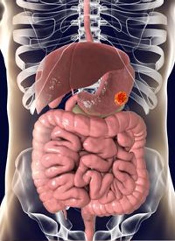
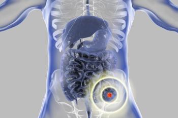
ROCKVILLE, Maryland-The FDA has approved a tiny ingestible video camera-the Given Diagnostic Imaging System (Given Imaging Ltd)-for use with other endoscopic and radiologic GI tract evaluations, to detect polyps, cancer, or causes of bleeding and anemia in the small intestine.
Advertisement
ROCKVILLE, MarylandThe FDA has approved a tiny ingestible video camerathe Given Diagnostic Imaging System (Given Imaging Ltd)for use with other endoscopic and radiologic GI tract evaluations, to detect polyps, cancer, or causes of bleeding and anemia in the small intestine.
The system includes a single-use, 11 × 30-mm capsule, known as the M2A, which contains a color video-imaging unit, light source, telemetry transmitter, and battery.
The patient swallows the capsule, which passes through the GI tract and is excreted naturally. A window at one end of the capsule allows the camera to take images of the intestine lining (2 pictures every second) for about 8 hours, long enough to view the entire small intestine but not the whole colon.
Images are sent to a data recorder worn around the patient’s waist and later transferred for viewing to a workstation equipped with the company’s application software.
Articles in this issue
over 24 years ago
High-Dose IL-2 Is Standard in Advanced Renal Cell Cancerover 24 years ago
RIT Safe, Effective in Elderly and Poor-Prognosis Patientsover 24 years ago
FDA Approves Xeloda/Taxotere Combination for Advanced Breast Cancerover 24 years ago
ODAC Recommends Approval of Radiolabeled Zevalinover 24 years ago
Proteomics Moves From the Laboratory to Clinical Researchover 24 years ago
Raltitrexed + Oxaliplatin for Advanced Colorectal Cancerover 24 years ago
Mental Fatigue Worries Chemotherapy Patientsover 24 years ago
Patients Urged to Work With Professionals Against Fatigueover 24 years ago
NCI Director Resigns to Head New Scientific InstituteAdvertisement
Related Content
Advertisement


Advertisement
Advertisement
Trending on CancerNetwork
1
Arlo-Cel Produces Deep Responses in Pretreated R/R Multiple Myeloma
2
FDA Clears Alpha DaRT Trial to Complete Enrollment in Recurrent GBM
3
Lurbinectedin Misses Survival End Point in Second-Line SCLC Trial
4
Belantamab Mafodotin Regimen Yields Enduring Responses in NDMM
5