|Articles|June 1, 2007
Oncology NEWS International
- Oncology NEWS International Vol 16 No 6
- Volume 16
- Issue 6
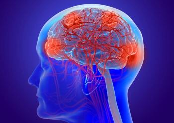
Intrathecal 131I-8H9 for CNS Mets
Radioimmunotherapy (RIT) with a radiolabeled monoclonal antibody 131I-8H9, delivered intrathecally, appears to have clinical utility when added to salvage therapy for patients with metastatic cancer to the central nervous system (CNS), according to a phase I study reported at the 2007 American Association of Cancer Research annual meeting (LB-4).
Advertisement
LOS ANGELESRadioimmunotherapy (RIT) with a radiolabeled monoclonal antibody 131I-8H9, delivered intrathecally, appears to have clinical utility when added to salvage therapy for patients with metastatic cancer to the central nervous system (CNS), according to a phase I study reported at the 2007 American Association of Cancer Research annual meeting (LB-4).
Disseminated malignant primary brain tumors and other cancers that metastasize through the cerebrospinal fluid (CSF) are challenging to treat. Antibody-based targeted therapies given through the CSF compartment have real therapeutic potential with regard to tumors that metastasize to the leptomeninges (LM), said principal investigator Kim Kramer, MD, of Memorial Sloan-Kettering Cancer Center, who presented the findings at a late-breaking trials session.
Monoclonal antibody 8H9 is a murine IgG1 antibody reactive with a wide spectrum of human solid tumors. The compound 131I-8H9, which is administered through a ventricular access device, has favorable pharmacokinetics in nonhuman primates and poses minimal toxicity.
The study involved 19 patients, aged 2 to 51 years, with refractory or recurrent CNS or LM malignancies that were shown to be 8H9 reactive. Multiple parenchymal lesions were observed in 7 patients and LM disease in 9. Tumors included neuroblastoma (n = 10), melanoma (n = 2), medulloblastoma (n = 3), pineoblastoma (n = 1), and ependymoma (n = 3).
Patients had baseline MRI of the brain and spine, and CSF cytology. Patients received a dosimetry dose of 2 mCi of intrathecal 131I-8H9, after which CSF and blood were sampled serially for pharmacokinetics, and nuclear scans were performed at 24 hours to evaluate drug localization. Therapeutic doses were 10, 20, 30, 40, and 50 mCi, with three to six patients per dose level. At 1 month post-injection, more MRI studies were done, and if no progressive disease or grade 3-4 toxicity was noted, the treatment was repeated.
The 19 patients received 56 injections: 16 patients received at least one full cycle (dosimetry and therapy injections), and 10 of the 16 received two full cycles; 3 received only the dosimetry dose. Mean dose to the CSF was 35.4 cGy/mCi, with 50% of injections being 20 to 50 cGy/mCi. Total CSF dose was 314 to 2,917 cGy. In contrast, the mean blood dose was 2.34 cGy/mCi, with an average CSF:blood ratio of 15:1 in all patients.
"So far, there has been a low toxicity profile, with no allergic, cardiovascular, or pulmonary toxicities," Dr. Kramer reported. "Transient grade 1-2 nausea and vomiting or fever was common in the immediate post-injection period, as was a transient rise in liver enzymes. Thus far, the maximum tolerated dose has not been reached, at 50 mCi."
Response Profile
Of the 16 patients receiving at least one full cycle, 8 had evaluable disease. In these patients, there was 1 complete response. Progressive disease was observed in 7 patients, 2 of whom remain alive with disease at more than 11 and 25 months since injection, Dr. Kramer reported.
She described the complete responder, a neuroblastoma patient who had multinodular leptomeningeal disease in the brain and spine with CSF positivity. This patient received one 30 mCi 131I-8H9 dose, and her scans no longer show disease, now more than 23 months since detection of the metastasis. This patient also had a rise in liver enzymes, but since she had a good response and this was her second relapse, Dr. Kramer received approval for two subsequent injections, and no additional toxicity was observed.
This patient also received the highest cGy/mCi estimates, suggesting that in patients with evaluable disease, there may be a dose-response relationship, Dr. Kramer said.
Eight of the 16 patients had no measurable disease, and all are alive in complete remission from 5 to 31+ months since treatment. "In contrast, we have previously shown that the CNS is a sanctuary site for neuroblastoma, and in our historical controls, the mean survival time, despite surgery, radiotherapy, and chemotherapy, for patients with relapsed neuroblastoma was approximately 6 months," she said.
Dr. Kramer now incorporates 131I-8H9 into a salvage regimen that includes gross total resection of disease, craniospinal irradiation with irinotecan (Camptosar) and temozolomide (Temodar), followed by intrathecal 131I-8H9. The possibility of systemic spread is addressed with intravenous administration of the 3F8 monoclonal antibody and oral temozolomide and isotretinoin (Accutane) treatment.
"We think the addition of radioimmunotherapy to CNS salvage regimens may improve survival. Thus far with this salvage regimen, 8 patients remain alive and in continuous remission 5 to 45 months since the time of CNS detection," she reported.
Articles in this issue
almost 19 years ago
ACS Launches Major Epidemiology Studyalmost 19 years ago
Pt Selection Key to Radioembolization of Liver Ca'salmost 19 years ago
Million Dollar Gotham Prize Announcedalmost 19 years ago
Diagnostic Dilemma: GI Diseasealmost 19 years ago
Velcade/Doxil Approved for Relapsed or Refractory Multiple Myeloma Ptsalmost 19 years ago
Junovan Fails to Win ODAC Nod for Osteosarcoma Treatmentalmost 19 years ago
Surveillance Colonoscopy Guidelines Not Being Followedalmost 19 years ago
Removing Stage IV Primary May Cut Mortalityalmost 19 years ago
Nuclear Export Inhibitors Testing Moving Forwardalmost 19 years ago
ODAC: orBec Yields No 'Substantial Efficacy' in GI GVHDAdvertisement
Related Content
Advertisement


Latest CME


Advertisement
Advertisement
Trending on CancerNetwork
1
Toxicity From CD19, BCMA Therapy May Inform B-Cell Malignancy Surveillance
2
Sac-TMT Significantly Improves PFS/OS in Pretreated Endometrial Cancer
3
Nogapendekin Alfa Plus BCG Exhibits Efficacy Advantage in NMIBC In Situ
4
Fianlimab Combo Does Not Significantly Improve PFS in Advanced Melanoma
5