
News|Articles|September 27, 2022
Kim Blenman, PhD, MS, Talks Development of Cell-Counting Software for Breast Cancer
Author(s)Nicholas Wrigley
Kim Blenman, PhD, MS, discussed the promising developments in cell-counting software for predicting pathologic complete response in breast cancer.
Advertisement
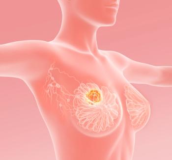
A study of image analysis–based software designed to algorithmically count tumor infiltrating lymphocytes (TILs) in the tumor microenvironments suggested that it’s at least as accurate as manual counting and may be predictive of treatment efficacy in breast cancer, according to results presented at the
“These types of software could be a very useful tool for pathologists to better enumerate different components within the tumor microenvironment,” Kim Blenman, PhD, MS, chief investigator of the study as well as Assistant Professor of Medicine (Medical Oncology) at Yale School of Medicine and Assistant Professor of Computer Science at the Yale School of Engineering and Applied Science in New Haven, Connecticut, said in an interview with CancerNetwork®.
QuPath software was used to test the quantifying capability of an analysis algorithm (CNN11) using data from the SWOG S0800 trial (NCT00856492) of neoadjuvant chemotherapy with or without bevacizumab (Avastin) in stage IIB to IIIC HER2-negative breast cancers. TILs quantification using QuPath open-source software and a convolutional neural network cell classifier (CNN11) yielded significantly higher pretreatment scores in cases showing pathologic complete response (pCR) vs residual disease (means, 31% vs 17%, P <.001). Additionally, the correlation between algorithmic and pathologist-read methods was high (r = 0.606, P <.0001), with areas under the prediction curve (AUC) of 0.709 and 0.627, respectively. Investigators thus concluded that image analysis–based TILs quantification displayed better outcome discrimination and predicted pCR better than pathologist-read stromal TILs quantification.
CancerNetwork®: What was the rationale for using image analysis–based TILs measurements as a predictor for complete response?
Blenman: Humans aren’t good at counting objects in large complex environments such as histology slides, and something we’ve learned over the past few years is that cell counts of certain objects such as TILs in histology slides actually matter. The [total] count of infiltrating immune cells, of actual cancer cells, and of accessory cells within the tumor microenvironment [affects] how the host’s immune system will remove the cancer cells. When we first tried to enumerate cell counts, we discovered that there are too many in the tumor microenvironment [for humans to easily arrive at a consistent and accurate count]. A solution to this problem is to design software which can count for us, and so we’ve designed a software algorithm (i.e., instructions) which can count individual cells that can differentiate between immune cells and tumor cells, and can estimate cell counts within certain components or certain tissue. [This software algorithm] is a great way to get those cell counts.
What were some of the key findings from this research?
This study aimed to validate a software algorithm that we had already used in melanoma. In this case, we wanted to see how it performed as a prognostic tool for treatments [in breast cancer]. We took advantage of a beautiful study from the Southwest Oncology Group, called SWOG S0800,2 which examined patients with stage IIB to IIIC HER2-negative breast cancer treated with and without bevacizumab. [That study found that] bevacizumab plus standard-of-care chemotherapy did increase patients’ chances of responding, but we wanted to [develop a tool] that could tease out which patients didn’t need to take that added bevacizumab. We embarked on this study using this particular algorithm to count cells because the [precise] TILs count could give us some idea of which patients will respond.
The normal way to find these cell counts is for pathologists to manually count through the oculars of the microscope. In this study, [we compared the manual method to] our software algorithm and found…the algorithm to be a bit more accurate. This is what we had anticipated, because humans [generally] estimate the total count rather than counting individually [to arrive at a precise number. By contrast], the software algorithm individually counts the cells which will naturally provide a better readout.
What do you feel is the multidisciplinary significance of this research?
This type of research opens many doors for our pathologists. [These types of software] can help investigators see things they could not normally see and it reduces the time it takes to do a robust job of [determining cell counts] and determine differences in immune cell populations. Additionally, we can use these tools in a health care environment to improve treatments for our patients.
Of course, there are still hurdles [to overcome] as we roll out these new tools. [For example], we need to understand how to implement these systems in a real-world setting and ensure that they aren’t designed only for high-resource environments. We have to ensure that these systems can be used in low-resource environments as well to help reduce the disparities in various communities and move closer to equity, especially here in the United States. Providing tools that are low-cost or or free to access would be accessible for any community [This is one of the ways we can] ensure we reduce disparities and increase equity.
Where might future research and development efforts go from here?
There is some controversy about using these types of tools, but the reality is that they are here to stay, and people are starting to use them. What we need to do is regulate them [to ensure] that their use does not harm patients. Many academic, government, and industry stakeholders have been coming together as a community to create some ground rules [regarding these tools] to ensure we do no harm. I am part of the steering and leadership council for a group called the Pathology Innovation Collaborative Community [PIcc], which brings together many stakeholders including faculty and staff from [schools like] Yale and Harvard, pharmaceutical companies, biotech companies, government agencies like the FDA and National Institutes of Health, as well as patients and patient advocates. The PIcc is a wonderful environment in which all stakeholders’ voices are heard. [We ask questions like]: what does everyone want to see in this tool? What are the basic things we need to see in any tools that use machine learning, or artificial intelligence, in any kind of patient care environment? We discuss what regulatory agencies, and what the field in general…feels should be the minimum standards [for new tools to meet]. [The PIcc] is based here in the United States, and is chaired by Jochen Lennerz, MD, PhD, an Associate Professor of Pathology at Harvard Medical School.
However, there are also international organizations. I am a member of the International Immuno-Oncology Biomarker Working Group on Breast Cancer [TILS Working Group], which is chaired by Roberto Salgado, MD, PhD; Sherene Loi, MD, PhD; and Carsten Denkert, MD. There are many ongoing clinical trials worldwide examining TILs as predictive or prognostic tools, and so there is a concerted effort to develop a nice cohort of leaders in the field, samples, patients, and patient advocates in this space to help extend the community of evaluators. I am also working with the FDA with Brandon Gallas, PhD to assemble a dataset of patient samples and statistical packages so that those developing algorithms and software tools for use in health care, [for example] in the context of TILs, will have a [ready-made] validation hurdle to determine if the tools they create are viable. We’re planning for these datasets [to be] freely available so that software developers can [easily] test their algorithms. Developing these robust statistical pipelines to standardize [how we validate these new Artificial Intelligence/Machine Learning tools] is not something we’ve ever done before as a field.
These systems are going to roll out soon. We are assembling training resources and other resources for people to examine these types of software tools.
What do you hope your colleagues took away from this research?
There is a place for everyone—whether they be academics, industry professionals, patients, or patient advocates—to be integrated in these developments. There are settings in which these new tools are being pushed forward and that are important for everyone to participate in. [It is important for a diverse group of people] to join these collaborative organizations so they can have a voice and better understand [these developments].
We are in an era of health care [which values collaboration]: everyone is working together to push things forward as a community, [including] clinicians, scientists, patients, and patient advocates.
[Lastly], we need to ensure we are designing these software tools so that people in low-income, low-resource environments can take advantage of them as well [rather than] only people in high-income environments. All of us in the global health community feel that this is an important way to reduce [socioeconomic] disparities and increase equity. We need to bring healthcare to the same standard of equity that [we want to see in broader] society.
Reference
Blenman K, Fanucci K, Bai Y, et al. Prediction of pathologic complete response to neoadjuvant chemotherapy in breast cancer (SWOG S0800) using image analysis-based tumor infiltrating lymphocyte measurements. J Clin Oncol. 2022;40(16):594. doi: 10.1200/JCO.2022.40.16_suppl.594
Advertisement


Advertisement
Advertisement
Trending on CancerNetwork
1
Analyzing ADC Options Across Metastatic TNBC
2
64 A Phase I/II Study of Pembrolizumab and Anti-CD3 x Anti-HER2 Armed Activated T-Cells in Metastatic Breast Cancer
3
Pembrolizumab Combo Shows Efficacy in Pediatric High-Risk Hodgkin Lymphoma
4
CD47 Antibody Combo Shows Meaningful Activity in Previously Untreated AML
5