
Press Release|Articles|January 15, 2026
Novel Theranostic Model Shows Curative Potential for Gastric and Pancreatic Tumors

Advertisement
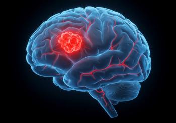
A newly developed radiopharmaceutical pair can precisely detect and effectively treat--completely eradicating tumors in certain preclinical models--gastric and pancreatic tumors. Targeting the well-defined and accessible biomarker claudin-18.2, the theranostic technique has the potential to move the field substantially closer to durable disease control and potentially cure--in otherwise difficult-to-treat solid tumors. This research was published in the January issue of The Journal of Nuclear Medicine.
Advanced upper gastrointestinal cancers, including esophageal, gastric, and pancreatic cancer, are among the deadliest cancers worldwide, accounting for one-third of all cancer deaths. Patients have very limited treatment options, especially in advanced stages, and current therapies provide only modest improvements in survival. As such, there is a critical unmet need for more effective and more precise approaches.
"Claudin-18.2 recently gained clinical attention following U.S. Food and Drug Administration approval of the gastric cancer treatment zolbetuximab, which targets the biomarker," said Shadi Esfahani, MD, MPH, nuclear medicine physician at Massachusetts General Hospital in Boston. "To further explore the utility of claudin-18.2, my colleagues and I developed a first-in-class claudin-18.2-targeted PET radiopharmaceutical and a therapeutic counterpart to identify and treat gastric and pancreatic tumors."
To evaluate the theranostic pair, preclinical murine models were developed with both pancreatic and gastric cancer cell lines. Serial PET imaging with 89Zr-DFO-zolbetuximab or 89Zr-DFO-IgG as a control was performed at one, three, and six days, followed by ex vivo analysis of biodistribution. Next, tumor-bearing mice received a single intravenous injection 177Lu-DOTA-zolbetuximab (high or low dose), non-radiolabeled zolbetuximab, 177Lu-DOTA-IgG, 177Lu-DOTA, or saline as a treatment. Effectiveness and toxicity of the treatments were evaluated by laboratory and histologic analyses.
Serial PET imaging demonstrated a high tumor uptake of 89Zr-DFO-zolbetuximab at all timepoints--significantly higher than the uptake seen in mice imaged with control 89Zr-DFO-IgG. High-dose 177Lu-DOTA-zolbetuximab resulted in reduced tumor growth in gastric and pancreatic mice models, with complete regression of most pancreatic tumors. No radiation-induced toxicities were observed during the study.
"Claudin 18.2 based theranostics could meaningfully change patient care in two important ways," noted Esfahani. "First, claudin 18.2 targeted PET imaging enables noninvasive identification of patients whose tumors strongly express this target. Second, claudin 18.2 targeted radiopharmaceutical therapy has the potential to deliver highly focused radiation directly to tumor cells, leading to significant tumor shrinkage and the possibility of improved survival."
She continued, "More broadly, the study underscores how advances in biomarker discovery, radiopharmaceutical development, and patient selection are bringing the field closer to treatments with curative potential rather than incremental benefit. As additional tumor-specific targets are identified and validated, molecular imaging-guided theranostics could increasingly enable personalized, highly effective, and safer cancer treatments, positioning nuclear medicine at the forefront of next-generation oncology."
Figure 2: Evaluation of [89Zr]Zr-DFO-zolbetuximab PET specificity in mouse models of GCa over multiple time points. (A and B) Maximum-intensity-projection serial PET/CT images for male nude mice implanted with GSU gastric tumors (white circles) imaged with [89Zr]Zr-DFO-zolbetuximab or [89Zr]Zr-DFO-IgG 1, 3, and 6 d after injection. (C and D) Quantitative in vivo biodistribution of [89Zr]Zr-DFO-zolbetuximab and [89Zr]Zr-DFO-IgG in tumor and selected tissues. ns = not significant on one-way ANOVA test.
The authors of "
Visit the
###
Please visit the
About JNM and the Society of Nuclear Medicine and Molecular Imaging
The Journal of Nuclear Medicine (JNM) is the world s leading nuclear medicine, molecular imaging and theranostics journal, accessed 15 million times each year by practitioners around the globe, providing them with the information they need to advance this rapidly expanding field. Current and past issues of The Journal of Nuclear Medicine can be found online at
JNM is published by the Society of Nuclear Medicine and Molecular Imaging (SNMMI), an international scientific and medical organization dedicated to advancing nuclear medicine, molecular imaging, and theranostics--precision medicine that allows diagnosis and treatment to be tailored to individual patients in order to achieve the best possible outcomes. For more information, visit
Advertisement
Related Content
Advertisement


Advertisement
Advertisement
Trending on CancerNetwork
1
FDA Approves Gedatolisib for HR+, HER2– PIK3CA Wild-Type Advanced Breast Cancer
2
FDA Clears Manufacturing Concern Behind Rivoceranib/Camrelizumab CRL in HCC
3
Sac-TMT Combo Meets PFS End Point in PD-L1–Negative NSCLC Trial
4
Gedatolisib Approval in HR+/HER2- Breast Cancer May Be “Practice Changing”
5