
|Articles|July 27, 2010
Nanoparticles reveal then kill cancers…maybe
Author(s)Greg Frieherr, Greg Frieherr
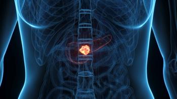
Go back to the beginning of MRI, in the early and mid-1980s, and you’ll find an almost rabid adoption of the modality, despite scant evidence of its clinical value. MRI has since done much to gain the trust of the medical community, opening a diagnostic cornucopia in the process. But the future has to bring more if MR is going to extend this legacy. Researchers at Wake Forest University Baptist Medical Center are working on it.
Advertisement
Go back to the beginning of MRI, in the early and mid-1980s, and you’ll find an almost rabid adoption of the modality, despite scant evidence of its clinical value. MRI has since done much to gain the trust of the medical community, opening a diagnostic cornucopia in the process. But the future has to bring more if MR is going to extend this legacy. Researchers at Wake Forest University Baptist Medical Center are working on it.
In research announced July 21 and
Multiwalled carbon
Iron-based imaging agents have been kicking around MRI labs for decades. What distinguishes them is the fact they absorb energy, laser energy to be exact, and turn it into heat.
Exposed to laser light while inside a tumor, these nanotubes become red-hot pokers that sear cancer at its cellular roots. Their iron content makes them visible with MRI, confirming they are in place to wreak oncologic mayhem.
Reflective of the research itself, progress with these nanoparticles is taking shape in tiny steps, actually mouse steps. As part of his ongoing Ph.D. thesis work at Wake Forest Baptist, Xuanfeng Ding demonstrated that the tiny particles were indeed visible under MRI and did heat up under laser light, so much that they could destroy tumor cells in which they have settled. Whether they ever will is hard to say.
A decade may pass from the inception of a good idea to the emergence of a pharmaceutical product. What’s remarkable-more remarkable than the potential of this one experimental concoction-is that this research is at once novel and routine: novel in the tailoring done to the particles to make them visible to MRI; routine in the tools applied during this process.
Nanotechnology is rapidly reshaping the future, it seems, for just about everything from imaging agents to electric car batteries. It is modern alchemy whose promise has not yet been achieved but, unlike medieval efforts to turn common metals into gold, likely will be.
How ironic that the very tiny will reshape the world on such a macro level.
Advertisement
Related Content
Advertisement


Advertisement
Advertisement
Trending on CancerNetwork
1
TOP Trial: Chemo-Osimertinib in TP53-Mutant EGFR-Mutated NSCLC
2
Five Gastrointestinal Cancer Abstracts You May Have Missed at ESMO GI 2026
3
How Are CELMoDs Reshaping Treatment in Multiple Myeloma?
4
Epcoritamab/R2 Earns European Approval for R/R Follicular Lymphoma
5