
|Articles|April 1, 1995
Oncology NEWS International
- Oncology NEWS International Vol 4 No 4
- Volume 4
- Issue 4
New Single-Cell Test May Prove Useful in Screening of Drugs
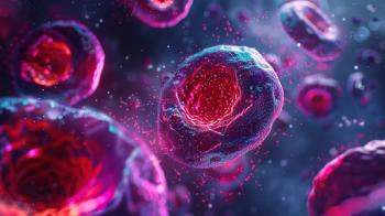
STANFORD, Calif--A new single-cell biological sensor system may someday allow rapid screening of cancer agents for biofunctional activity (see illustration on page 1). The test could be used, for example, to identify compounds that bind to or block receptors for biomolecules such as growth factors or other cytokines, or to highlight potentially harmful metabolites.
Advertisement
STANFORD, Calif--A new single-cell biological sensor system maysomeday allow rapid screening of cancer agents for biofunctionalactivity (see illustration on page 1). The test could be used,for example, to identify compounds that bind to or block receptorsfor biomolecules such as growth factors or other cytokines, orto highlight potentially harmful metabolites.
Richard N. Zare, PhD, who headed the research in the Departmentof Chemistry at Stanford University, told Oncology News Internationalthat the technique may have an "important impact in cancerresearch, depending on how well we develop a library of receptorsthat we can place on cells."
Cells with known receptors could be used as biosensors to screencompounds for the presence of the ligand that binds to that receptor,he said.
The technique uses the responses of single cells to detect individualbiomolecules separated from complex chemical mixtures by capillaryelectrophoresis. This method solves a past problem with biologicalsensors in that a cell can respond to many different biologicalcompounds. "So when a cell reacts to something in a mixture,it is difficult to identify just what the cell is responding to.Capillary electrophoresis allows us to identify the specific compoundsthat are triggering the reaction," Dr. Zare said.
How It Works
The separation of a compound takes place in a fused silica capillarywith an inside diameter of 25 microns, a fraction of the sizeof a human hair. Each type of molecule moves through the liquidin the capillary at a different rate, depending on its electricalcharge, size, shape, and other factors.
When a separated biomolecule reaches the end of the capillary(a process that takes between 5 and 30 minutes), it is directedonto the single-cell biosensor, positioned on a microscope. Inthis way, a tiny amount of a given chemical can be delivered toan individual cell.
The new system was able to detect individual components of a mixtureof acetylcholine, bradykinin, and adenosine triphosphate witha sensitivity that ranged from a few million molecules to a fewthousand (Science 267:74-76, 1995), levels comparable to thoseachieved by the best detection techniques currently available,Dr. Zare said.
The molecules were identified through their binding to receptorson a cultured rat brain cell (PC-12 cell). Binding triggered therelease of intracellular calcium, which was detected by a fluorescentdye.
The researchers also showed that it may prove feasible to usegenetic engineering to make biosensors for use in detecting specificcompounds. Unfertilized frog eggs were microinjected with a messengerRNA that encoded a serotonin receptor. The altered eggs boundserotonin to the membrane surface, whereas unaltered ones didnot. This experiment used a technique that involved measurementof transmembrane currents rather than fluorescence.
Dr. Zare's co-workers in the study were Jason B. Shear, currentlyat Cornell, and Harvey A. Fishman, Delia Garigan, Nancy L. Allbritton,and Richard H. Scheller, all of Stanford.
Articles in this issue
about 31 years ago
First Results of Avicidin Trialsabout 31 years ago
Chemo Patients Often Develop Menstrual Irregularitiesabout 31 years ago
Growth Factor Receptor Blockade Moving From Laboratory to Clinicabout 31 years ago
Controlled-Release AZT Gets FDA Go-Ahead for New Drug Investigationabout 31 years ago
Vinorelbine Plus Chemo Promising in Advanced Diseaseabout 31 years ago
Bone Substudy a Part of Tamoxifen Prevention Trialabout 31 years ago
Ultrasound Breast Screens Useful in Selected Womenabout 31 years ago
New Roles Forecast for Endocrine Therapyabout 31 years ago
Antisense Drug Against BCL-2 Effective in Animal Cancer Modelsabout 31 years ago
Anticancer Drugs From Zeneca in Regulatory Phase of DevelopmentAdvertisement
Related Content
Advertisement


Advertisement
Advertisement
Trending on CancerNetwork
1
From Conference to Practice: Top 5 Takeaways From EHA 2026
2
Emerging T-Cell Engagers and Novel Immunotargets in Multiple Myeloma
3
Givastomig Earns FDA Fast Track Designation in HER2-Negative Gastric Cancer
4
Evaluating Bladder-Sparing Strategies and Key GU Data From ASCO 2026
5