
|Articles|March 1, 2007
Oncology NEWS International
- Oncology NEWS International Vol 16 No 3
- Volume 16
- Issue 3
Diagnostic Dilemma: GI Disease
Author(s)Matthew J. Mckinley, MD
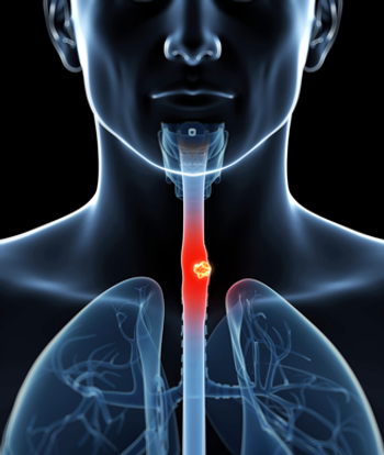
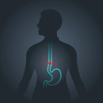
Barrett's esophagus and documented high-grade dysplasia (HGD)
Advertisement
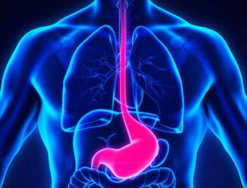
Test your diagnostic skills with the following endoscopic quiz.
This 76-year-old man is being followed closely for Barrett's esophagus and documented high-grade dysplasia (HGD). The HGD involved the esophagus in a diffuse manner. Photodynamic therapy (PDT) was delivered 8 months ago after a full discussion of the therapeutic options and risks. Due to multiple medical conditions, the patient was assessed as being a high surgical risk for esophagectomy. There is no history of dysphagia, abdominal pain, or weight loss. He has a long history of heartburn that is well controlled on medications.
His past history is significant for stable angina, hypertension, mild chronic obstructive pulmonary disease (COPD), diverticulosis, and a small stable abdominal aortic aneurysm.
His medical therapy consists of high-dose proton pump inhibition and cardiac medications. There is a long history of prior smoking, and he consumes alcohol rarely. Family history is negative for malignancy and positive for heart disease.
Physical examination is unremarkable. Laboratory evaluation is also normal.
The patient has undergone repeat upper gastrointestinal endoscopies since the photodynamic therapy. Photographs were obtained during these examinations.
1) Photograph A reveals:
a) An esophageal mass
b) Evidence of residual Barrett's epithelium
c) Duodenal ulceration
d) A mid-esophageal diverticulum
e) An area of suspicious columnar lining
2) Appropriate management at this time includes:
a) Laparoscopic fundoplication
b) Surgical resection
c) Random four-quadrant biopsies every 1 to 2 cm and biopsies of abnormal areas
d) Empiric treatment for H pylori
e) Selective vagotomy
3) Photograph B demonstrates:
a) An erythematous polyp
b) A visible vessel
c) An aorto-enteric fistula
d) Endoscopic mucosal resection (EMR)
e) Endometriosis
1. The answers are (b) evidence of residual Barrett's epithelium and (e) an area of suspicious columnar lining. In endoscopic photograph A, there is no evidence of mass, ulceration, or diverticulum. Between the 3 and 6 o'clock positions in photo A, there is evidence of squamous re-epithelialization in areas previously lined with columnar epithelium. The remaining mucosa represents residual Barrett's lining. The photograph also demonstrates a patch of suspicious mucosa in the upper portion of the photograph, especially between the 11 and 12 o'clock positions.
2. The answer is (c) random four-quadrant biopsies every 1 to 2 cm and biopsies of abnormal areas. At this point, there is no indication for laparoscopic fundoplication, surgical resection, empiric treatment for H pylori, or selective vagotomy. Multiple biopsies are most appropriate in a search for persistent high-grade dysplasia and occult carcinoma. Biopsies revealed areas of squamous re-epithelialization, columnar lining with areas of intestinal metaplasia, and focal HGD in the proximal segment of Barrett's where the abnormal patch of mucosa was visualized.
3. The answers are (a) an erythematous polyp and (d) endoscopic mucosal resection (EMR). The image is not consistent with a visible vessel or aorto-enteric fistula, and endometriosis would be extremely unusual in the esophagus, especially in a male!
The erythematous polyp is actually created by suctioning mucosa into a cap located on the tip of the endoscope and applying a rubber band. The "polyp" is then removed by snare and cautery. The snare can be seen in the photograph.
The advantage of EMR over ablative techniques such as photodynamic therapy (PDT) is that the tissue can be recovered and examined microscopically. With ablation, a larger surface area can be treated, but the tissue is destroyed and is not available for examination. Initially, the patient had a large surface area with diffuse involvement with HGD; therefore, PDT was selected. Subsequently, he only had evidence of focal HGD, making EMR an attractive therapeutic option.
Besides PDT, other ablative techniques include multipolar electrocoagulation, argon plasma coagulation, and Yag Laser. Bipolar radiofrequency and cryotherapy have recently been introduced as alternative methods. Reports in the literature have suggested excellent long-term results with PDT and EMR in properly selected patients. As this case shows, a combination of treatment options can best suit an individual patient's needs.
Selected Readings
Ell C et al: Endoscopic mucosal resection of early cancer and high-grade dysplasia in Barrett's esophagus. Gastroenterology 118:670-677, 2000.
Overholt BF et al: Photodynamic therapy for Barrett's esophagus with dysplasia and/or early stage carcinoma: Long-term results. Gastrointest Endosc 58:183-188, 2003.
Articles in this issue
almost 19 years ago
Peptide-Based Breast Ca Vaccines Promising in Early Trialsalmost 19 years ago
Imatinib Responses in CML May Take Timealmost 19 years ago
IV Vidaza Approved; Oral Formulation to Be Testedalmost 19 years ago
Study Confirms Avastin Advantage in Advanced NSCLCalmost 19 years ago
Legal Services Should Be a Component of Standard Cancer Carealmost 19 years ago
Mouse Virus Evidence Suggests Viral Basis for Breast Caalmost 19 years ago
Groups Oppose Ruling on Access to Experimental Drugsalmost 19 years ago
Satellite Allows Digital Mammography Screening for Rural Native Americansalmost 19 years ago
Nexavar Effective in Advanced HCC: Phase III Trial Stoppedalmost 19 years ago
H&N Ca Patients With CR to CRT May Not Need SurgeryNewsletter
Stay up to date on recent advances in the multidisciplinary approach to cancer.
Advertisement
Related Content
Advertisement
Latest CME


Advertisement
Advertisement
Trending on CancerNetwork
1
Modifiable Risk Factors Suggest Potential for Improving Cancer Prevention
2
2026 Tandem Meetings: What’s the Latest Research in Multiple Myeloma?
3
Dato-DXd Receives Priority Review in Unresectable/Metastatic TNBC
4
Barriers to CAR T-Cell Referral and Center Access in Multiple Myeloma
5