
|Articles|January 1, 2006
Oncology NEWS International
- Oncology NEWS International Vol 15 No 1
- Volume 15
- Issue 1
3D Digital Breast Tomosynthesis Provides Clearer Picture of Breast Anatomy Than Does Standard Mammography
Based on their initial experience with 3D digital breast tomosynthesis, radiologists at Dartmouth Hitchcock Medical Center and Dartmouth Medical School believe the investigational technique "is closer to the truth than standard mammography," Steven Poplack, MD, associate professor of diagnostic radiology and obstetrics and gynecology, said at the 2005 meeting of the Radiological Society of North America (abstract SSG01-05).
Advertisement
CHICAGO-Based on their initial experience with 3D digital breast tomosynthesis, radiologists at Dartmouth Hitchcock Medical Center and Dartmouth Medical School believe the investigational technique "is closer to the truth than standard mammography," Steven Poplack, MD, associate professor of diagnostic radiology and obstetrics and gynecology, said at the 2005 meeting of the Radiological Society of North America (abstract SSG01-05).
Describing one of five cases of malignancy in a study of 98 women who underwent digital breast tomosynthesis after an abnormal mammogram, Dr. Poplack noted that conventional mammograms showed an area of asymmetry with architectural distortion, which is consistent with a suspicious mass. The corresponding tomosynthesis images showed the abnormality in much greater detail, including the shape and the anatomical features at the margins of the lesion, which allowed the radiologists to characterize the mass as almost certainly an invasive breast cancer.
The tomosynthesis image also revealed a darker gray area within the tumor, which is an unusual finding in a breast malignancy and is commonly associated with fat. The pathological slide of the tumor confirmed the presence of fat surrounded by malignant cells.
"Tomosynthesis better reflects what is really going on in the breast than conventional mammography is able to do. That will hopefully translate into more accurate diagnosis," Dr. Poplack said.
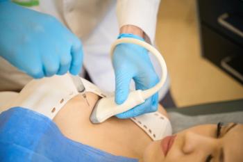
With standard mammography as well as digital breast tomosynthesis, the breast is placed under compression and exposed to x-rays. The difference with 3D tomosynthesis is that the x-ray tube moves across the breast in an arc, and the machine takes a series of low-dose miniexposures. Data from a tomosynthesis examination are reconstructed into thin sections, which may be displayed in individual single scans or as a fly-through movie of image after image.
The study, sponsored by Hologic, Inc., included women who had abnormal digital screening mammograms and required additional diagnostic work-up: 98 women were included, and 99 breasts were examined. As many as three tomosynthesis views were obtained on each woman, corresponding to the views that had been obtained with diagnostic mammograms. Tomosynthesis images were reviewed prospectively by one of two breast imaging specialists, and the images were compared with diagnostic mammograms.
Superior 33% of the Time
The radiologists concluded that tomosynthesis was superior to mammography 33% of the time, equivalent 54% of the time, and inferior 12% of the time. With regard to inferior findings, Dr. Poplack explained, "It wasn't as though tomosynthesis didn't see the abnormality. It just didn't characterize it as well." The presence of calcifications accounted for 9 (69%) of the 13 cases for which mammography was superior to tomosynthesis.
Tomosynthesis also decreased the rate of recall for screening mammography by 40%. In one case, a 1-cm area of asymmetry with ill-defined margins on standard screening mammography was recommended for biopsy. However, the tomosynthesis images taken in the same projection as the mammography scans clearly showed fibroglandular tissue.
"If tomosynthesis had been included in the screening study, I don't think the patient would have been recalled for additional evaluation. Had the tomosynthesis been included as a diagnostic work-up, the patient would not have been recommended for biopsy," he said.
Both tomosynthesis and conventional mammography detected the same number of breast cancers. Of a total of five cases of malignancy, one was identified incidentally with ultrasound. Although this malignancy was visible in retrospect with tomosynthesis, it remained occult on conventional mammography.
When benign lesions were present, tomosynthesis depicted the abnormalities much more definitively than conventional mammography, Dr. Poplack said. "So I believe that not only are we going to get a benefit in the detection of breast abnormalities, we also will decrease the rate of false negatives," he said.
He cautioned that the study was small and therefore does not have the statistical power to validate the effectiveness of digital breast tomosynthesis for diagnosis or for screening. He nevertheless predicted that tomosynthesis will be quickly validated as a diagnostic tool that is equivalent to mammography. Because of the number of patients and the number of cancers that must be evaluated in breast cancer screening trials, he stated that the adoption of digital breast tomosynthesis in screening would be slower.
Articles in this issue
over 20 years ago
FDA Approves Nexavar for Use in Advanced Kidney Cancerover 20 years ago
Avastin Benefits Metastatic Breast Cancerover 20 years ago
Amgen to Acquire Abgenixover 20 years ago
Better Prognosis for ‘Elsewhere' Local Breast Ca Recurrencesover 20 years ago
Herceptin/Taxotere Ups DFS in Early HER2+ Breast Caover 20 years ago
NIH 2006 Budget Is Likely to Be Lower Than in 2005over 20 years ago
Amooranin, a Plant Compound, Shows Potential as Cancer Treatmentover 20 years ago
FDA Launches 7 Initiatives With European Drug RegulatorsAdvertisement
Related Content
Advertisement


Advertisement
Advertisement
Trending on CancerNetwork
1
FDA Accepts NDA for Daraxonrasib in Pretreated Metastatic Pancreatic Cancer
2
RASolute 302 and the Future of Pancreatic Cancer Treatment: Paul Oberstein, MD, on the KRAS Breakthrough
3
Mapping the Full CAR T-Cell Journey in DLBCL From Referral to Survivorship
4
Balancing Oncologic Outcomes With Patient Priorities in Prostate Cancer Care
5