News|Articles|December 16, 2019
- ONCOLOGY Vol 33 No 12
- Volume 33
- Issue 12
Hypercalcemia in a Patient with Small Lymphocytic Lymphoma/Chronic Lymphocytic Leukemia
Advertisement
THE CASE
A 64-year-old white man was originally diagnosed with chronic lymphocytic leukemia (CLL)/small lymphocytic lymphoma (SLL) back in 2007. Initial presentation was with fatigue, elevated white blood cell counts with lymphocytosis, and a palpable lymph node in his neck. A biopsy of the neck node showed lymphoid infiltrates consistent with B-cell CLL that was later confirmed by flow cytometry. At initial presentation, he had favorable prognostic indicators including mutated immunoglobulin heavy chain gene (IgHV), negative zeta-associated protein kinase70 kDa expression, and deletion of 13q14. He was managed with a watch-and-wait strategy until 2013 when he developed anemia (hemoglobin: 9.8 g/dL), mild thrombocytopenia (platelet count 116,000 per cubic mL) and an elevated white blood cell (WBC) count with lymphocytosis (WBC: 61,000 per cubic mL, with 79% lymphocytes). He was treated with 6 cycles of fludarabine, cyclophosphamide, and rituximab (Rituxan)1 with a complete clinical response. In 2015, work up for some respiratory symptoms discovered a 6 cm by 2.8 cm left upper lung mass. This turned out to be a T3 N0 M0 squamous cell non–small cell lung cancer (NSCLC) that was treated with curative intent left upper lobectomy followed by adjuvant chemotherapy with cisplatin and vinorelbine. In 2017, he presented with tiredness, axillary lymphadenopathy, anemia, elevated WBCs with lymphocytosis and thrombocytopenia. He was treated with 6 cycles of bendamustine and rituximab2 resulting in normalization of blood counts and a decrease in axillary, supraclavicular, and mediastinal lymphadenopathy which lasted 1 year.
In 2018, he presented with anorexia, vomiting, mild mental confusion, and weight loss. Further work up revealed hypercalcemia (Ca: 17.6 mg/dL, normal: 8.1-10.1 mg/dL), elevated WBC with lymphocytosis (WBC: 19.7 per cubic mL, with 81% lymphocytes), mild anemia (hemoglobin: 12.4 g/dL) and thrombocytopenia (platelet count 86,000 per cubic mL). Serum level of parathyroid hormone (PTH) was suppressed (PTH intact: 10 pg/mL, normal 12-65 pg/mL). The serum level of PTH-related peptide (PTHrP) was slightly elevated (PTHrP: 2.5 pmol/L, normal <2 pmol/L). The serum levels of vitamin D (25-hydroxyvitamin D: 25.6 ng/dL, normal: 30-61 ng/dL) and 1,25-dihydroxyvitamin D (1,25-dihydroxyvitamin D: 8.4 pg/mL, normal: 19.9-79.3 pg/mL) were both low. Serum thyroid-stimulating hormone and thyroxin levels were normal. Ultrasound of thyroid and parathyroid glands and a bone scan were normal. Computed tomography (CT) scans revealed enlarged lymph nodes in the bilateral neck, supraclavicular, mediastinum, hilum, central mesentery, aortocaval, and bilateral groin areas (Figure 1).
Which of the following statements is/are true?
A. This clinical picture is consistent with the recurrence of squamous cell NSCLC with paraneoplastic hypercalcemia.
B. This clinical picture is consistent with the recurrence of CLL/SLL with paraneoplastic hypercalcemia.
C. This clinical picture is consistent with the recurrence of CLL/SLL with Richter`s transformation leading to hypercalcemia.
D. All of the above statements are true.
CORRECT ANSWER (B): This clinical picture is consistent with the recurrence of CLL/SLL with paraneoplastic hypercalcemia.
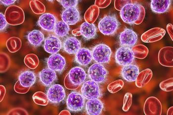
ANSWER EXPLAINED: Bronchoscopy and biopsy of the subcarinal lymph node showed atypical lymphoid infiltrates consistent with CLL. Bone marrow aspiration and a biopsy revealed hypercellular bone marrow with CLL (Figure 2), and flow cytometry and cytogenetic evaluation revealed high CD38 expression and deletion of 17p. General treatment measures for hypercalcemia (intravenous hydration, furosemide, and bisphosphonates) were not effective until the start of ibrutinib (Imbruvica) and obinutuzumab (Gazyva).3 After 4 cycles of this regimen, the patient had a complete hematologic response, normalization of calcium levels, and a partial radiologic response to his lymphadenopathy.
Outcome of This Case
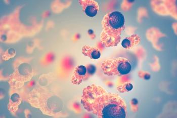
Six months into his continued treatment with ibrutinib, the patient developed mild mental confusion, anorexia, and abdominal pain. Further evaluation revealed an elevated serum calcium level of 17.3 mg/dL, and CT scans revealed a mild to moderate increase in neck and chest lymphadenopathy and a large increase in abdomen and pelvis lymphadenopathy (Figure 3). A biopsy of a para-aortic node showed lymphoid tissue with B-cell CLL/SLL (Figure 4). The majority of cells in the biopsy specimen were small to intermediate in size with some scattered large cells. There were several areas with prolymphocytes and paraimmunoblasts consistent with proliferation centers. He was started on venetoclax (Venclexta)4 and had a short-lived clinical response lasting 1 month. Coincidentally, with disease progression, hypercalcemia reappeared. A bone marrow biopsy showed hypercellular bone marrow with atypical lymphoid proliferation with large cells concerning for Richter’s transformation (Figure 5). He was evaluated for chimeric antigen receptor T-cell therapy; however, his underlying disease progressed rapidly. The patient and family decided on hospice care. His overall condition deteriorated with persistent refractory hypercalcemia and he passed away shortly thereafter.
Key Points
• Hypercalcemia of malignancy is a relatively frequent finding in patients with cancer affecting nearly half of patients, particularly in those with advanced stage cancer and limited survival of several months. It is not clear whether this poor prognosis is related to the advanced stage of malignancy or if hypercalcemia is simply a marker of more aggressive cancer.
• Hypercalcemia in B-cell CLL/SLL is extremely rare. Proposed mechanisms for hypercalcemia include concurrent primary hyperparathyroidism, secondary hyperparathyroidism related to tumor production of vitamin D (1,25- dihydroxycholecalciferol) or PTHrP, or secondary osteolysis by an osteoclast-activating factor such as TNF-alpha.
• Hypercalcemia of B-cell CLL/SLL has also been observed in patients with transformation of CLL to prolymphocytic leukemia or large-cell (immunoblastic) lymphoma (Richter's syndrome). The presumed pathogenesis of hypercalcemia is the release of serum cytokines TNF-alpha and IL-6, which are thought to increase bone resorption.
Discussion
Here we present a patient with 2 distinct primary cancers, CLL/SLL and squamous cell NSCLC, who developed rather refractory hypercalcemia that eventually led to his death.
The differential diagnosis of hypercalcemia includes multiple pathologic entities, but the 2 most common etiologies include primary hyperparathyroidism and hypercalcemia of malignancy.5 The estimated yearly prevalence of hypercalcemia for all cancers is 1nearly 3%.6 Hypercalcemia is 4 times more common in stage IV cancer and it is associated with a poor prognosis. The most common cancers presenting with malignant hypercalcemia are lung cancer, multiple myeloma, and renal cell carcinoma. These are followed by breast and colorectal cancers, and the lowest rates are reported in prostate cancer.6 Hypercalcemia of malignancy is a relatively frequent finding in patients with cancer, affecting almost hald of patients,7,8 particularly in those with advanced stage cancer.9 Patients with hypercalcemia of malignancy tend to have limited survival of several months, and it is not clear whether this poor prognosis is related to the advanced stage of malignancy or that hypercalcemia is simply a marker of more aggressive cancer.10
Among several major mechanisms leading to malignancy-related hypercalcemia are excessive tumor production of PTHrP, osteolytic metastatic disease, ectopic activity of 1-alpha-hydroxylase coupled with the production of 1,25-dihydroxycholecalciferol, the excessive production of PTH by parathyroid carcinoma and extra parathyroid malignancies, and hypercalcemia due to paraprotein binding. Approximately 80% of malignancy-related hypercalcemia is mediated by the production of PTHrP.9 PTHrP is normally produced and secreted by various cells, including normal breast cells, and participates in the development of various tissues.11 PTHrP acts on osteoblasts, leading to enhanced synthesis of receptor activator of nuclear factor-kappa B ligand, with subsequent activation of osteoclasts and bone resorption with calcium release into the blood stream and increased renal calcium reabsorption. Squamous cell cancers, urinary tract cancers (renal cancer and bladder cancer), breast cancer, non-Hodgkin lymphoma, and ovarian cancer account for the majority of malignancies leading to hypercalcemia through PTHrP.9
Hypercalcemia develops in 6 % of patients with lung cancer. Squamous cell carcinoma is the most frequent type of lung cancer in patients with hypercalcemia. In patients with lung cancer, hypercalcemia is more commonly caused by tumor secretion of PTHrP.12,13 Our patient had an early stage (T3N0M0) squamous NSCLC that was treated with curative intent. Radiologic findings, bronchoscopy, and biopsy of a subcarinal lymph node all supported the diagnosis of recurrent CLL/SLL rather than recurrent lung cancer.
Hypercalcemia in B-cell CLL is extremely rare, with less than 30 cases reported in the English literature in the past 50 years.14,15-17 In a retrospective study of 1200 patients with B-cell CLL, only 7 patients (0.006%) were found to have high calcium levels.18 Proposed mechanisms for hypercalcemia in patients with CLL include concurrent primary hyperparathyroidism, secondary hyperparathyroidism related to tumor production of vitamin D (1,25-dihydroxycholecalciferol) or PTHrP, or secondary osteolysis by an osteoclast-activating factor such as tumor necrosis factor-alpha (TNF-alpha).10 The mechanism of dysregulated production of vitamin D (1,25-dihydroxycholecalciferol) has been postulated to be similar to that of hypercalcemia due to sarcoidosis; however, it has not been clarified if the primary source of 1,25-dihydroxycholecalciferol is leukemia cells or surrounding macrophages in this setting.19 Hypercalcemia of CLL has also been observed in patients with transformation of CLL to prolymphocytic leukemia or large-cell (immunoblastic) lymphoma (Richter’s syndrome).20,21 The presumed pathogenesis of hypercalcemia is the release of serum cytokines TNF-alpha and interleukin 6 (IL-6), which are thought to increase bone resorption.22 It has also been reported that hypercalcemia in CLL can occur without any of the above mechanisms.23
In our case, the low serum PTH, low PTHrP, and normal vitamin D (1,25-dihydroxycholecalciferol) levels, without any other solid tumor, support that hypercalcemia was not the result of a primary or secondary hyperparathyroid state. It is also unlikely that the hypercalcemia in our patient was secondary to osteolysis due to lack of extensive bony involvement. While the initial presentation with hypercalcemia showed no signs of Richter’s transformation, later in the course of his disease, the patient became refractory to treatment and a bone marrow sample at that point confirmed large-cell transformation with Richter`s syndrome.
Here, we present a very rare case of B-cell CLL/SLL in a patient who presented with symptomatic hypercalcemia. The patient initially responded to treatment but progressed within 6 months and his disease transformed into Richter`s syndrome with a dismal outcome. Future research on the pathogenesis of hypercalcemia in CLL will increase our understanding of this rare complication and potentially help discover appropriate management options for these patients.
Financial Disclosure: The authors have no significant financial interest in or other relationship with the manufacturer of any product or provider of any service mentioned in this article.
FIVE KEY REFERENCES
1. Thompson PA, Tam CS, O'Brien SM, et al. Fludarabine, cyclophosphamide, and rituximab treatment achieves long-term disease-free survival in IGHV-mutated chronic lymphocytic leukemia. Blood. 2016;127:303-309. doi: 10.1182/blood-2015-09-667675.
2. Cuneo A, Follows G, Rigolin GM, et al; GIMEMA, European Research Initiative on CLL (ERIC) and UK CLL forum. Efficacy of bendamustine and rituximab as first salvage treatment in chronic lymphocytic leukemia and indirect comparison with ibrutinib: a GIMEMA, ERIC and UK CLL FORUM study. Haematologica. 2018;103(7):1209-1217. doi: 10.3324/haematol.2018.189837.
3. Moreno C, Greil R, Demirkan F, et al. Ibrutinib plus obinutuzumab versus chlorambucil plus obinutuzumab in first-line treatment of chronic lymphocytic leukaemia (iLLUMINATE): a multicentre, randomised, open-label, phase 3 trial. Lancet Oncol. 2019;20(1):43-56. doi: 10.1016/S1470-2045(18)30788-5.
4. Korycka-Wolowiec A, Wolowiec D, Kubiak-Mlonka A, Robak T. Venetoclax in the treatment of chronic lymphocytic leukemia. Expert Opin Drug Metab Toxicol. 2019;15(5):353-366. doi: 10.1080/17425255.2019.1606211.
5. Endres DB. Investigation of hypercalcemia. Clin Biochem. 2012;45(12):954-963. doi: 10.1016/j.clinbiochem.2012.04.025.
ABOUT THE SERIES EDITORS:
Maria T. Bourlon, MD is Associate Professor, Head Urologic Oncology Clinic, National Researcher. Instituto Nacional de Ciencias Médicas y Nutrición Salvador Zubirán. Mexico City, Mexico. She is also a Member of ASCO’s IDEA Working Group.
E. David Crawford, MD, is Chairman, Prostate Conditions Education Council;
Editor in Chief, Grand Rounds in Urology; and Professor of Urology, University of California San Diego, La Jolla, California.
References:
1. Thompson PA, Tam CS, O'Brien SM, et al. Fludarabine, cyclophosphamide, and rituximab treatment achieves long-term disease-free survival in IGHV-mutated chronic lymphocytic leukemia. Blood. 2016;127:303-309. doi: 10.1182/blood-2015-09-667675.
2. Cuneo A, Follows G, Rigolin GM, et al; GIMEMA, European Research Initiative on CLL (ERIC) and UK CLL forum. Efficacy of bendamustine and rituximab as first salvage treatment in chronic lymphocytic leukemia and indirect comparison with ibrutinib: a GIMEMA, ERIC and UK CLL FORUM study. Haematologica. 2018;103(7):1209-1217. doi: 10.3324/haematol.2018.189837.
3. Moreno C, Greil R, Demirkan F, et al. Ibrutinib plus obinutuzumab versus chlorambucil plus obinutuzumab in first-line treatment of chronic lymphocytic leukaemia (iLLUMINATE): a multicentre, randomised, open-label, phase 3 trial. Lancet Oncol. 2019;20(1):43-56. doi: 10.1016/S1470-2045(18)30788-5.
4. Korycka-Wolowiec A, Wolowiec D, Kubiak-Mlonka A, Robak T. Venetoclax in the treatment of chronic lymphocytic leukemia. Expert Opin Drug Metab Toxicol. 2019;15(5):353-366. doi: 10.1080/17425255.2019.1606211.
5. Endres DB. Investigation of hypercalcemia. Clin Biochem. 2012;45(12):954-963. doi: 10.1016/j.clinbiochem.2012.04.025.
6. Jick S, Li L, Gastanaga VM, Liede A. Prevalence of hypercalcemia of malignancy among cancer patients in the UK: analysis of the Clinical Practice Research Datalink database. Cancer Epidemiol. 2015;39(6):901-907. doi: 10.1016/j.canep.2015.10.012.
7. Burt ME, Brennan MF. Incidence of hypercalcemia and malignant neoplasm. Arch Surg. 1980;115(6):704-707.
8. Vassilopoulou-Sellin R, Newman BM, Taylor SH, Guinee VF. Incidence of hypercalcemia in patients with malignancy referred to a comprehensive cancer center. Cancer. 1993;71(4):1309-1312.
9. Stewart AF. Clinical practice. Hypercalcemia associated with cancer. N Engl J Med. 2005; 352(4):373-379.
10. Ralston SH, Gallacher SJ, Patel U, Campbell J, Boyle IT. Cancer-associated hypercalcemia: morbidity and mortality. Clinical experience in 126 treated patients. Ann Intern Med. 1990;112(7):499-504.
11. Wysolmerski JJ. Parathyroid hormone-related protein: an update. J Clin Endocrinol Metab. 2012;97(9):2947-2956. doi: 10.1210/jc.2012-2142.
12. Hiraki A, Ueoka H, Takata I, et al. Hypercalcemia-leukocytosis syndrome associated with lung cancer. Lung Cancer. 2004;43(3):301-307.
13. Thomas L, Kwok Y, Edelman MJ. Management of paraneoplastic syndromes in lung cancer. Curr Treat Opt Oncol. 2004;5(1):51-62.
14. Briones J, Cervantes F, Montserrat E, Rozman C. Hypercalcemia in a patient with chronic lymphocytic leukemia evolving into Richter's syndrome. Leuk Lymphoma. 1996;21(5-6):521-523.
15. Salahudeen AA, Gupta A, Jones JC, Cowan RW, Vusirikala M, Kwong C, Naina HV. PTHrP-induced refractory malignant hypercalcemia in a patient with chronic lymphocytic leukemia responding to denosumab. Clin Lymphoma Myeloma Leuk. 2015;15(9): e137-e140. doi: 10.1016/j.clml.2015.06.007.
16. Kampfenkel T, Baraniskin A, Teschendorf C, Schmiegel W, Massenkeil G. A rare case of chronic lymphocytic leukemia with hypercalcemia induced by elevated parathyroid hormone-related peptides. Acta Haematol. 2010;124(1):57-60. doi: 10.1159/000314646.
17. Vlasveld LT, Pauwels P, Ermens AA, Aarnoudse WH, Ooms HW, Haak HR. Parathyroid hormone-related protein (PTH-rP)-associated hypercalcemia in a patient with an atypical chronic lymphocytic leukemia. Neth J Med. 1999;54(1): 21-26.
18. Fain O, el M'Selmi A, Dosquet C, et al. Hypercalcemia in B cell chronic lymphocytic leukaemia. Br J Haematol. 1994;87(4):856-858.
19. Seymour JF, Gagel RF. Calcitriol: the major humoral mediator of hypercalcemia in Hodgkin's disease and non-Hodgkin's lymphomas. Blood. 1993;82(5):1383-1394.
20. Harousseau JL, Flandrin G, Tricot G, Brouet JC, Seligmann M, Bernard J. Malignant lymphoma supervening in chronic lymphocytic leukemia and related disorders. Ritcher's syndrome: a study of 25 cases. Cancer. 1981;48(6):1302-1308.
21. Beaudreuil J, Lortholary O, Martin A, et al. Hypercalcemia may indicate Richter's syndrome: report of four cases and review. Cancer. 1997;79(6):1211-1215.
22. Mirrakhimov AE. Hypercalcemia of malignancy: an update on pathogenesis and management. N Am J Med Sci. 2015;7(11):483-493. doi: 10.4103/1947-2714.170600.
23. Macintyre EA. Hypercalcaemia in chronic lymphatic leukaemia. Postgrad Med J. 1986;62(727):393-394.
Articles in this issue
over 6 years ago
Understanding Lung Cancer Treatment Advancesover 6 years ago
Evaluating the Role of Targeted Therapy in Lung Cancerover 6 years ago
The Future of CAR T-Cell TherapyAdvertisement
Related Content
Advertisement


Advertisement
Advertisement
Trending on CancerNetwork
1
TOP Trial: Chemo-Osimertinib in TP53-Mutant EGFR-Mutated NSCLC
2
How Are CELMoDs Reshaping Treatment in Multiple Myeloma?
3
Five Gastrointestinal Cancer Abstracts You May Have Missed at ESMO GI 2026
4
FLAURA2: Osimertinib Plus Chemo in EGFR-Mutant NSCLC
5