


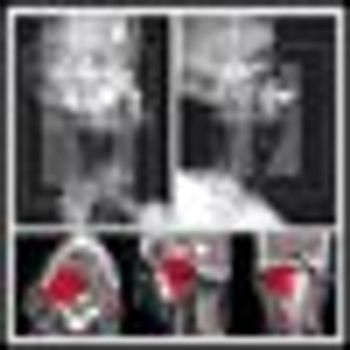
In their article, Rusthoven and colleagueshighlight the utility ofcombined positron-emission tomography/computed tomography(PET-CT) imaging for diagnosing primaryand recurrent head and neckcarcinoma, and for defining tumor targetvolumes for radiotherapy treatmentplanning in the head and neck. PEToffers noninvasive measures of tumorbiology yet suffers from limited spatialresolution; the physiologic informationobtained with PET is complementaryto the high-resolution structural informationobtained with CT or magneticresonance imaging (MRI).
Head & Neck Cancer
Latest News
Advertisement
Advertisement
Positron-emission tomography(PET) and computed tomography(CT) fusion imaging is arapidly evolving technique that is usefulin the staging of non–small-celllung cancer (NSCLC), Hodgkin’s disease,ovarian cancer, gastrointestinalstromal tumors, gynecologic malignancies,colorectal malignancies,and breast cancer. In their article,Rusthoven et al[1] describe the roleof PET-CT in head and neck malignanciesand include a review of allcurrently available literature. Accordingto the authors, PET-CT is usefulfor staging head and neck carcinomasand for target volume delineation duringradiation treatment planning.
The fusion of 18-fluorodeoxyglucose (FDG) positron-emission tomography(PET) with computed tomography (CT) offers both anatomicand physiologic delineation of head and neck cancers. PET-CT is usefulin the staging of head and neck carcinomas and may identify unsuspecteddistant metastasis that may alter treatment. PET-CT may alsohelp in target volume delineation during radiotherapy (RT) treatmentplanning. Better characterization of the target may improve local controlas well as spare normal tissues from RT sequelae.
In 2005, it is estimated that head and neck cancers will comprise 2%-3% of allcancers in the United States and account for 1%-2% of all cancer deaths. Thistotal includes 20,780 cases of oral cavity cancer, 9,880 cases of laryngeal cancer,and 8,590 cases of pharyngeal cancer. Most patients with head and neckcancer have metastatic disease at the time of diagnosis (regional nodal involvementin 43% and distant metastasis in 10%).
The past several years have seenthe fruition of a new era in cancertherapy-targeted approachesusing biologic modifiers.However, as the clinical experiencewith novel inhibitors grows, some ofthe premises on which the treatmentswere designed are being challenged,and clinical findings are leading to newparadigms. Drs. Song and Raben providea forward-thinking review of thestatus of epidermal growth factor receptor(EGFR)-targeted therapy in headand neck cancer, a paper that serves toboth highlight progress and raise issuesthat continue to challenge the implementationof targeted therapy.
The treatment of head and neck cancer has been at the forefront ofnovel therapeutic paradigms. The introduction of drugs that interactwith selective biologic pathways in the cancer cell has generated considerableattention recently. A wide variety of new compounds that attemptto target growth-signaling pathways have been introduced intothe clinic. A majority of studies in the clinic have focused on epidermalgrowth factor receptor (EGFR) antagonists, but future studies will likelybuild upon or complement this strategy with agents that target angiogenicor cell-cycle pathways. EGFR activation promotes a multitude ofimportant signaling pathways associated with cancer development andprogression, and importantly, resistance to radiation. Since radiationtherapy plays an integral role in managing head and neck squamouscell cancer (HNSCC), inhibiting the EGFR pathway might improveour efforts at cancer cure. The challenge now is to understand whenthe application of these EGFR inhibitors is relevant to an individualpatient and how or when these drugs should be combined with radiationor chemotherapy. Are there molecular markers available to determinewho will respond to EGFR inhibitors and who should be treatedwith alternative approaches? What are the mechanisms behind intrinsicor acquired resistance to targeted agents, and how do we preventthis problem? We need to formulate integrated laboratory/clinicalresearch programs that address these important issues.
Head and neck squamous cellcarcinoma (HNSCC) is themost common malignant neoplasmarising in the upper aerodigestivetract, accounting for approximately40,000 new cases each yearin the United States. Despite increasingawareness of the importance ofearly cancer detection, the majorityof patients continue to present withadvanced-stage (stage III/IV) disease.Standard therapy has included surgicalresection followed by externalbeamradiation or chemotherapy inconjunction with radiotherapy(chemoradiation). Although no prospectiveclinical trials have comparedsurgical with nonsurgical therapies,only 50% of patients are cured of theirprimary tumors. Even with successfuleradication of the primary tumor,second primary tumors can be expectedto occur at the rate of 4% to 5% peryear and are often fatal. Given the extrememorbidity and mortality ofHNSCC, new and innovative treatmentsbased on the biologic alterations thatcharacterize these tumors are required.
NEW ORLEANS-Cetuximab (Erbitux) plus high-dose radiation therapy (RT) significantly improved survival in patients with advanced head and neck cancer, compared with RT alone, according to results of an international phase III trial reported at the 40th Annual Meeting of the American Society of Clinical Oncology (abstract 5507).
The 14 reports in this special supplement discuss theuse of the cytoprotectant amifostine in patients withcancer of the head and neck, esophagus, lung, andcervix, as well as those with lymphoma and acutemyelogenous leukemia. Discussions focus on thepotential of this agent to both reduce radiation sideeffects such as xerostomia and permit doseescalation of chemotherapy and/or radiotherapy.Improvements in treatment outcome and quality oflife as a result of cytoprotection are examined.
The 14 reports in this special supplement discuss theuse of the cytoprotectant amifostine in patients withcancer of the head and neck, esophagus, lung, andcervix, as well as those with lymphoma and acutemyelogenous leukemia. Discussions focus on thepotential of this agent to both reduce radiation sideeffects such as xerostomia and permit doseescalation of chemotherapy and/or radiotherapy.Improvements in treatment outcome and quality oflife as a result of cytoprotection are examined.
In this issue of ONCOLOGY, Kutleret al eloquently address the concept,application, and controversiesof a planned neck dissection inpatients with head and neck carcinomaand nodal metastasis who receivenonsurgical therapy to the primary tumor.As stated lucidly in the article,planned neck dissection arose in thehistorical context of low rates of completeresponse in patients with N2/3neck disease treated with conventionallyfractionated radiotherapy, coupledwith low surgical salvage ratesamong patients who failed in the neck.Hence, the concept evolved that allpatients with N2/3 neck disease shouldundergo a planned neck dissection regardlessof response to radiotherapy.
The presence of regional nodal metastases represents a significantadverse prognostic factor for patients with squamous cell carcinoma ofthe head and neck. Early-stage head and neck cancers, localized to theprimary site without regional lymph node metastases have excellentcure rates with either surgery or radiation therapy. The presence ofregional metastases results in cure rates that are approximately half ofthose obtainable in early-stage disease. Therefore, due to the significantadverse impact of neck metastases on prognosis, the treatment ofthe neck remains a vital part of the decision-making process in determiningtherapy for head and neck cancer.
The recent recognition that theaddition of concurrent chemotherapyto definitive radiationcan improve locoregional control, organpreservation, and survival in patientswith squamous cell head andneck cancer has had a significant impacton our management choices.Chemoradiotherapy data from metaanalyses,cooperative group trials, andlarge tertiary care institutions now suggestthat there is a realistic potentialfor cure in almost all patients withlocoregionally confined disease, and thefocus has increasingly shifted towardthe impact of our treatments on longtermfunction. In the past, control ofneck node involvement often requireda comprehensive neck dissection, a procedureassociated with some degree oflong-term morbidity. In this review,Kutler, Patel, and Shah address the importantquestion of whether the neckdissection should be a planned componentin the management of patientstreated with definitive concurrentchemoradiotherapy.
COPENHAGEN, Denmark-Radiation in 6 fractions per week is significantly better than the same dose given on a more leisurely 5-fractions-per-week schedule for treating squamous-cell head and neck cancer, according to investigators from the Danish Head and Neck Cancer Study Group (DAHANCA).
Oropharyngeal mucositis hasbeen reported as the mostbothersome side effect by patientsundergoing myeloablative regimens,and it remains a therapy-limitingtoxicity of radiation and chemotherapyfor head and neck cancer. JoelEpstein and Mark Schubert providean informative review of progressmade over more than a decade of researchon the pathophysiology andmanagement of oropharyngeal mucositisin patients undergoing cancertreatment.
Rash is a class effect of HER1/epidermal growth factor receptor(EGFR)-targeted agents, and has occurred with high frequency and ina dose-dependent manner in clinical trials of these agents in cancerpatients. Analysis of phase II trials of erlotinib (Tarceva) in non–smallcelllung cancer, head and neck cancer, and ovarian cancer shows asignificant association between rash severity and objective tumor response.Rash severity was highly significantly associated with survivalin patients with non–small-cell lung cancer receiving erlotinib; mediansurvival in patients with no rash was 46.5 days, compared with257 days in those with grade 1 rash (P < .0001) and 597 days in thosewith grade 2/3 rash (P < .0001). Similarly, for the combined non–smallcelllung cancer, head and neck cancer, and ovarian cancer studies,median survival in patients with no rash was 103 days, compared with191 days in those with grade 1 rash (P = .0001) and 266 days in thosewith grade 2/3/4 rash (P = .0001). Similar findings have been madewith cetuximab (Erbitux) and in some settings with gefitinib (Iressa).The strong association of rash severity with response/survival suggeststhat rash may serve as a marker of response to erlotinib treatment andmay be used to guide treatment to obtain optimal response. Dosingerlotinib at the maximum tolerated dose, which is associated with morefrequent and more severe rash, may improve response rates and survivaldurations. Further study of the potentially important associationbetween rash and outcome of treatment with EGFR-targeted agents isneeded.
Paragangliomas are unusual tumorsof the head and neck butshould be included in the differentialdiagnosis of lateral neck masses.Although malignant paragangliomasare possible, these tumors are usuallybenign. Nevertheless, treatment canlead to great morbidity and possiblemortality. The article by Drs. Hu andPersky addresses a multidisciplinaryapproach to these lesions.
Paragangliomas most commonly occur in the carotid body, jugulotympanicarea, and vagus nerve but have also been reported in otherareas of the head and neck. These tumors are highly vascular andcharacteristically have early blood vessel and neural involvement,making their treatment particularly challenging. Surgery has traditionallybeen the preferred method of treatment, especially in light of recentadvances in technique. However, compared to radiation therapy, it canresult in a higher incidence of cranial nerve dysfunction. Radiationtherapy has the advantage of avoiding the increased morbidity ofsurgery while offering an equal possibility of cure. Part 2 of this articlediscusses radiation therapy as primary treatment of patients who areineligible for surgery and the elderly and infirm. Results with radiotherapyare comparable to those achieved with surgery. The efficacy ofsalvage therapy with either surgery or radiation is discussed, and atreatment algorithm for these tumors is proposed.
We have reviewed with interestthe article by Drs. Huand Persky and would liketo congratulate them on an excellentand comprehensive overview of theevaluation and management ofparagangliomas of the head and neck.Their review begins with an excellentlydetailed description of thedisease and staging work-up. Withmodern imaging, most paragangliomasare convincingly diagnosed basedon typical location (carotid bifurcation,nodose ganglia of the vagusnerve, middle ear along tympanic plexus,or near jugular bulb) and characteristicradiographic appearance(hypervascular, intensely enhancingmass). A tissue diagnosis is usuallyunnecessary for such lesions.
Paragangliomas most commonly occur in the carotid body, jugulotympanicarea, and vagus nerve but have also been reported in otherareas of the head and neck. These tumors are highly vascular andcharacteristically have early blood vessel and neural involvement,making their treatment particularly challenging. Surgery has traditionallybeen the preferred method of treatment, especially in light of recentadvances in technique. However, compared to radiation therapy, it canresult in a higher incidence of cranial nerve dysfunction. Radiationtherapy has the advantage of avoiding the increased morbidity ofsurgery while offering an equal possibility of cure. Part 1 of this two-partarticle focuses on techniques for diagnosing paraganglioma and theindications for and use of surgery as primary treatment. The complicationscommonly associated with surgery are reviewed, and strategies forrehabilitation of affected patients are presented.
Part of the multidisciplinary approach to cancer care involves surgical intervention. This is harmoniously interwoven through the efforts of the surgical oncologist and the reconstructive surgeon. As elegantly pointed out by Drs. Hasen, Few, and Fine, the reconstructive surgeon’s role in the management of malignancy is critical, involving the restoration of form and function. Sometimes, as in breast reconstruction, quality of life is improved by the restoration of form; other times, as in head and neck reconstruction, it is improved by the restoration of form and function. In fact, due to the significant morbidity associated with major ablation of head and neck cancer, such radical surgery would not be feasible without concomitant reconstruction.
Quon and Harrison have performed a considerable service to patients with head and neck cancers by reminding the oncology community that a state-of-the-art treatment team must include state-of-the-art brachytherapy
Drs. Quon and Harrison have written an excellent review on the role of brachytherapy in the management of head and neck cancer. Brachytherapy is a time-honored technique, and the authors have carefully reviewed the pertinent literature extolling its virtues. However, there are many papers that fail to document efficacy of brachytherapy over conventional techniques, demonstrating that, similar to surgery, the technique is both patient- and operator-dependent.
Brachytherapy is a therapeutic modality that may provide a significant improvement in the therapeutic ratio when appropriately applied, and hence, is an appealing treatment strategy for the head and neck. For several
Flavopiridol [2-(2-chlorophenyl 5 ,7-dihydroxy-8-[cis-(3-hydroxy-1-methyl-4-piperidinyl)-4H-1-benzopyran-4-one, hydrochloride] is a semisynthetic flavone with a novel structure compared with that of polyhydroxylated flavones, such as quercetin and genistein.[1] It is derived from rohitukine, an alkaloid isolated from the stem bark of Dysoxylum binectariferum, a plant indigenous to India.[2] Originally synthesized and supplied by Hoechst India Limited, flavopiridol is provided to the Division of Cancer Treatment and Diagnosis of the National Cancer Institute (NCI) by Aventis Pharmaceuticals, Inc.
Advertisement
Advertisement
Trending on CancerNetwork
1
FDA Approves Afami-cel in Metastatic Synovial Sarcoma
2
The Moonlight Shift: Dr Joshua Richter on Myeloma, Myths, and the C-Word
3
Current Treatment Landscape of HER2-Positive Metastatic Breast Cancer and Phase 3 Trial Overview
4
How Are Key Lymphoma Data From ASCO and EHA 2026 Shaping Treatment?
5